Showing posts with label cardiology. Show all posts

Wednesday, April 11, 2012

what's a TAVI?


vck8sm5h Transcatheter Aortic Valve Implantation (TAVI) Reduces Mortality Rate Compared to Standard Therapy


RAGING HYPOTHETICAL:
--Hypotensive, elderly gentleman presents to your ED, records show he's had a TAVI

--You nod your head with a reassurring look, then run to google...

--Next time you won't need to - because you read this post.


WHAT'S A TAVI?

Transcatheter Aortic-Valve Implantation (TAVI) for patients with severe aortic stenosis who are not candidates for surgery.

What it looks like:

kdaesp1huv Transcatheter Aortic Valve Implantation (TAVI) Reduces Mortality Rate Compared to Standard Therapy


How it is placed: (so cool and worth watching)
The implantation procedure involves accessing a femoral artery, performing balloon valvuloplasty, then advancing the device across the native valve. During rapid right ventricular pacing, a balloon is inflated to deploy the valve and the frame.

Video animation: http://youtu.be/_eiI6S62zOs


Evidence it works:

At one year, the rate of death from any cause was 30% with TAVI vs. 50.7% with standard treatment (balloon aortic valvuloplasty and or medical therapy)


Not so good: TAVI had a higher incidence of strokes and major vascular complications compared to standard treatment, however this was included in rate of death.


Now you know :)


Submitted by S. Morris.

Tuesday, March 27, 2012

chest pain with recent negative stress test

EP MONTHLY EXCERPT:
--nice overview on stress tests from March 2011(see reference link)


DOES THE RECENT NEGATIVE STRESS MEAN THIS PATIENT CAN'T HAVE ACS?
--No.
--stress tests are good, not great


SOME ROUGH EXAMPLES:
--Sensitivity for single vessel CAD:
  • exercise ECG stress test ~68%
  • exercise nuclear stress test (thallium) 60-82%
  • exercise nuclear stress test (sestamibi) 82-97%
  • adenosine/dipyridamole nuclear stress 77-92%
  • dobutamine nuclear stress 80-100%

BOTTOM LINE:
--stress sensitivity is not perfect
--negative stress test does NOT rule out ACS


Reference(s): ep monthly article, picture

Tuesday, March 20, 2012

the TASER-ed patient

PHYSIOLOGY OF TASERS:
--nice article in Emergency Medicine News from Feb '12 (link below)


HIGHLIGHTS:
--TASERs deliver electrical current to cause diffuse muscular contraction, thus incapacitating

--people who need to be TASERed may be drugged up, overexherted, or sustain trauma, so there are other things to think about

--in studies with healthy subjects, there were minimal (returning to baseline in 10 minutes) or no changes in pulse, 02 saturation, bicarb, lactate, electrolytes, troponin, EKGs, acidosis


EVERYTHING'S RELATIVE:
--TASER joule output: 0.36-1.76 joules


RANDOM FACT:
--TASER stands for "Thomas A. Swift's electric rifle", after the developer's childhood hero


BOTTOM LINE:
--asymptomatic, awake post-TASERed patient, unlikely to need routine labs/monitoring


Submitted by S. Lee.


Reference(s): EMN article, review article, taser joules, picture

Tuesday, February 7, 2012

cardiac tamponade and electrical alternans

POSSIBLE EKG FINDINGS IN CARDIAC TAMPONADE:
--sinus tachycardia
--low voltage
--electrical alternans


ELECTRICAL ALTERNANS:
--beat to beat alternation in QRS appearance
--related to back & forth swinging motion of the heart in the pericardial fluid (thought this was a nice way of picturing it)


Reference(s): uptodate.com: cardiac tamponadependulum, EKG

Tuesday, January 24, 2012

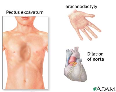
where do patient’s with Marfan Syndrome dissect?

AORTIC DISSECTION & MARFAN SYNDROME:
--The major cardiovascular manifestation in Marfan Syndrome is a progressive dilatation of the ascending aorta, leading to aortic aneurysm formation and eventually to fatal aortic rupture or dissection. Aortic dissection in early adult life is the leading cause of death.

--The ascending and descending aorta are both abnormal in Marfan Syndrome.

· The descending aorta is affected in two out of three patients during aortic dissection, and is the site of most complications which occur during follow-up.

· Aortic dissection limited to the descending aorta can occur in patients without dilatation of the ascending aorta.

--Dissection of the descending aorta was associated with dissection of ascending aorta in 43% and was isolated in 20% of cases.


BOTTOM LINE:
--dissections in Marfan involve descending aorta ~2/3 of the time, but these frequently involve the ascending aorta also
--if you have a patient with Marfan, and are worried about a dissection...worry about both (ascending/descending)


Submitted by J. Gullo.


Reference(s): PMID: 20232788, medscape article, picture

Friday, January 20, 2012

elevated troponin: what if its not an MI?

QUICK REVIEW:
--Myocardial necrosis indicated by elevated troponin is NOT always due to atherosclerotic CAD

--Troponin has high sensitivity for detecting very small amount of myocardial cell death

--Troponin is released in the blood due to irreversible as well as reversible cell damage AND does ≠ permanent myocyte damage


EXPANDED DIFFERENTIAL:
--Demand ischemia: sepsis/SIRS, hypotension, hypovolemia, SVT/afib, LVH

--Myocardial ischemia: coronary vasospasm, ICH/stroke, ingestion of sympathomimetic agents

--Direct myocardial damage: cardiac contusion, ICD shock, cardiac infiltrative d/o (amyloidosis), chemotherapy, myocarditis/pericarditis, heart transplant

--Myocardial strain: CHF, PE, PHTN, COPD, strenuous exercise

--Chronic renal insufficiency


Submitted by F. DiFranco.


Reference(s): Jeremias A. & Gibson M. Narrative review: alternative causes for elevated cardiac troponin levels when acute coronary syndromes are excluded. Annals of Internal Medicine. 142(9):786-791, May 2005.

Thursday, December 29, 2011

PE and the ECG

STUDY:
--retrospective chart review, 130 patients with diagnosed PEs, 140 controls with negative CTPAs
--checked 15 different ECG parameters between the groups, to see if people with PEs had particular ECG findings that normals didn't


NOTABLE NUMBERS:
--Sinus tachycardia (found in 39% of PE group vs. 24% of control group, P <0.01)
--S1 Q3 T3 pattern (12% vs. 3%, P <0.01)
--atrial tachyarrhythmias (15% vs. 4%, P <0.005)
--Q wave in lead III (40% vs. 26%, P <0.02)
--Q3 T3 pattern (8% vs. 1%, P <0.02)


NICE RECAP:
--"We conclude that 1) standard 12-lead ECG findings can increase the pretest probability of pulmonary embolism before performing CT pulmonary angiography; and that 2) the ECG findings have relatively low likelihood ratios to have clinical use."


Reference(s): study, picture

Tuesday, December 6, 2011

hypotension and valvular dysfunction: chicken or the egg?

CASE PARTICULARS:
--SOB, hypotension
--labs notable for slightly elevated troponin, AKI


DIAGNOSTICS:
--could not undergo CTPA to r/o PE due to AKI
--ECHO in ED shows no RV dilation but did show severe MR and TR.
--pt intubated secondary to massive fluid resuscitation

--ECHO following day was normal. 
--Once rehydrated, pt did much better.  No PE was found on CTPA.

WHAT HAPPENED?
--hypovolemia exacerbated systolic anterior motion of mitral valve causing severe dynamic MR and TR, causing cardiogenic shock. 

TEACHING POINTS:
--This patient had LV outflow tract obstruction by a hyperdynamic anterior motion of the mitral valve leaflets during systole, causing a severe mitral regurgitation due to the blockage of the outflow tract as well as opening of the mitral valve during systole. 
--This mimics HOCM, but was in a structurally normal heart. 
--Obstructive physiology, and especially systolic anterior motion of the mitral valve, can be caused by various disorders including hypercontractile states such as hypovolemia, anemia, beta agonist drugs, D-transposition of the great arteries, congenital/acquired abnormalities of the mitral valve/papillary muscles, and immediately after aortic valve surgery for aortic stenosis (due to acute afterload reduction).

10-SECOND TAKEAWAY:
--suspect massive PE, can’t to CTPA, think of bedside echo to look for RV dilation/strain
--severe hypercontractile states (e.g. hypovolemia) can result in obstructive physiology, including valvular dysfunction
--can be transient, should improve with fluids (good idea for treatment)

Submitted by T. Boyd.

Refence(s): Rosen B et al. “Hypovolemia-Induced Reversible Severe Mitral Regurgitation Due to Left Ventricular Outflow Tract Obstruction.” Echocardiography. 19:8; Nov. 2002.