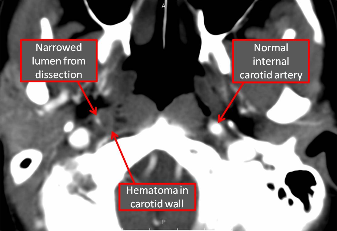
CAROTID (AND VERTEBRAL) DISSECTIONS: QUICK HITS
--first consult call to Neurologists (less commonly Vascular Surgeons)
--Vascular imaging is a must, but CTA is more
sensitive than MRA.
--To catch on MRI,
specific sequences are required so check with your Neurologist/Radiologist to
figure out which.
--Dissections that
extend intracranially have a high incidence of forming SAH, especially when
heparin is started, thus making heparin have higher mortality than
anti-platelet drugs.
--Extracranial
dissections are usually treated with antiplatelet drugs and anticoagulation.
Submitted by T. Boyd.
Reference(s): Shea et al. Carotid and Vertebral Artery Dissections in the Emergency Department. Emergency Medicine Practice.14;4. April 2012; picture
Showing posts with label neurology. Show all posts
Wednesday, May 23, 2012
Monday, May 7, 2012
epileptic vs. non-epileptic seizure: what are the signs?
WHAT ARE THE SIGNS?
--review by Avbersek & Sisodiya in J Neurol Neurosurg Psychiatry (link below)
--looked at 34 studies
--tried to tease out what signs distinguish epileptic vs. non-epileptic seizure
WHAT DID THEY FIND?
--signs that favor psychogenic non-epileptic seizures (specificity)
--signs that favor epileptic seizures (specificity)
--the sensitivities for all of these signs were horrible, so can't use them to rule anything out
--sometimes excluded frontal lobe partial seizures
--insufficient evidence: gradual onset, flailing/thrasing, opisthotonus, tongue biting, urinary incontinence
Reference(s): review article, picture
--review by Avbersek & Sisodiya in J Neurol Neurosurg Psychiatry (link below)
--looked at 34 studies
--tried to tease out what signs distinguish epileptic vs. non-epileptic seizure
WHAT DID THEY FIND?
--signs that favor psychogenic non-epileptic seizures (specificity)
- fluctuating course (96%)
- asynchronous movements (93-96%)
- pelvic thrusting (96-100%)
- side-to-side head/body movement (96-100%)
- closed eyes (74-100%)
- ictal crying (100%)
- memory recall (96%)
--signs that favor epileptic seizures (specificity)
- occurance from sleep (100%)
- post-ictal confusion (88%)
- stertorous (snoring) breathing (100%)
--the sensitivities for all of these signs were horrible, so can't use them to rule anything out
--sometimes excluded frontal lobe partial seizures
--insufficient evidence: gradual onset, flailing/thrasing, opisthotonus, tongue biting, urinary incontinence
Reference(s): review article, picture
Thursday, April 26, 2012
Spontaneous Cervical and Intracranial Arterial Dissections
(scroll to bottom for quick hits)
Background
-Arterial dissections can cause stroke in both young in old populations, and is the most common cause of stroke in the young
-Extracranial dissection is more common than intracranial dissection
-Stroke is generally caused by either diminished blood flow from intraluminal thrombus or embolized clot
Pathogenesis (stick with it)
-Dissection are believed to begin with a tear in the media of the vessel wall, leading to bleeding in the wall itself
-partially coagulated intramural blood can enter the lumen which activates platelets and
the coagulation cascade leading to intraluminal thrombus.
-Intramural blood can accumulate leading to compression of the lumen
Risk Factors
-Connective tissue and vascular disorders are thought to be main risk factors:
(e.g. Ehlers-Danlos Syndrome (type IV), Marfan Syndrome, Polycystic kidney disease, cystic medial necrosis, Fibromuscular dysplasia)
-many are caused by trauma
Diagnosis
-based on radiologic criteria taken from the Strategies Against Stroke Study for Young Adults in Japan (SASSY) using CTA or MRI/MRA
-Primary rules are based on finding an intimal flap or double lumen on Angiogram or MRI/MRA, or repeated non-specific findings associated with dissection on multiple studies.
Symptoms
-Head or neck pain
-Horner Syndrome - occurs when the sympathetic fibers on the ICA are stretched. It is usually just partial with ptosis and miosis but no anhidrosis.
Difference between Carotid and Vertebral Dissections
-Transient monocular blindness occurred only with internal carotid dissection
-Ischemic stroke is more common in vertebral dissections
-Neck pain and recent minor cervical trauma are more common in vertebral dissections
-Proportion of men and a recent infection are more common in ICA
Treatment
-Antithrombotic therapy (antiplatelet or anticoagulation) is the primary initial treatment for ischemic stroke and TIA caused by arterial dissection
-For intracranial dissection, antiplatelet therapy is often the treatment of choice
-For extracranial dissection, anticoagulation initially followed by 6 months of warfarin therapy as opposed to antiplatet therapy is often chosen
-Endovascular and surgical therapy are generally only reserved for recurrent ischemic events
BOTTOM LINE:
-arterial dissection is more common cause of stroke in the young
-extracranial (vertebral, carotid) more common than intracranial dissection
-sx: headache, neck pain, stroke-like symptoms, Horner's syndrome (for ICA)
-risk factors: connective tissue disorders, trauma
-dx: angiography, MRI/MRA
-tx: antithrombotics/anticoagulation (aspirin, heparin, coumadin, etc.); surgical/endovascular for recurrent events
Submitted by J. Grover.
Reference(s): Caplan, LR and Biousse V. “Cervicocranial Artery Dissections.” J Neuro-Opthalmol. 2004; 24:299-305. Maruyama, H et al. “Spontaneous Cervicocephalic Arterial Dissection with Headache and Neck Pain as the Only Symptom.” J Headache Pain (2012) 13: 247-253. “Spontaneous Cerebral and Cervical Artery Dissection: Treatment and Prognosis”. Uptodate. “Spontaneous Cerebral and Cervical Artery Dissection: Clinical Features and Diagnosis”. Uptodate., picture
Background
-Arterial dissections can cause stroke in both young in old populations, and is the most common cause of stroke in the young
-Extracranial dissection is more common than intracranial dissection
-Stroke is generally caused by either diminished blood flow from intraluminal thrombus or embolized clot
Pathogenesis (stick with it)
-Dissection are believed to begin with a tear in the media of the vessel wall, leading to bleeding in the wall itself
-partially coagulated intramural blood can enter the lumen which activates platelets and
the coagulation cascade leading to intraluminal thrombus.
-Intramural blood can accumulate leading to compression of the lumen
Risk Factors
-Connective tissue and vascular disorders are thought to be main risk factors:
(e.g. Ehlers-Danlos Syndrome (type IV), Marfan Syndrome, Polycystic kidney disease, cystic medial necrosis, Fibromuscular dysplasia)
-many are caused by trauma
Diagnosis
-based on radiologic criteria taken from the Strategies Against Stroke Study for Young Adults in Japan (SASSY) using CTA or MRI/MRA
-Primary rules are based on finding an intimal flap or double lumen on Angiogram or MRI/MRA, or repeated non-specific findings associated with dissection on multiple studies.
Symptoms
-Head or neck pain
-Horner Syndrome - occurs when the sympathetic fibers on the ICA are stretched. It is usually just partial with ptosis and miosis but no anhidrosis.
Difference between Carotid and Vertebral Dissections
-Transient monocular blindness occurred only with internal carotid dissection
-Ischemic stroke is more common in vertebral dissections
-Neck pain and recent minor cervical trauma are more common in vertebral dissections
-Proportion of men and a recent infection are more common in ICA
Treatment
-Antithrombotic therapy (antiplatelet or anticoagulation) is the primary initial treatment for ischemic stroke and TIA caused by arterial dissection
-For intracranial dissection, antiplatelet therapy is often the treatment of choice
-For extracranial dissection, anticoagulation initially followed by 6 months of warfarin therapy as opposed to antiplatet therapy is often chosen
-Endovascular and surgical therapy are generally only reserved for recurrent ischemic events
BOTTOM LINE:
-arterial dissection is more common cause of stroke in the young
-extracranial (vertebral, carotid) more common than intracranial dissection
-sx: headache, neck pain, stroke-like symptoms, Horner's syndrome (for ICA)
-risk factors: connective tissue disorders, trauma
-dx: angiography, MRI/MRA
-tx: antithrombotics/anticoagulation (aspirin, heparin, coumadin, etc.); surgical/endovascular for recurrent events
Submitted by J. Grover.
Reference(s): Caplan, LR and Biousse V. “Cervicocranial Artery Dissections.” J Neuro-Opthalmol. 2004; 24:299-305. Maruyama, H et al. “Spontaneous Cervicocephalic Arterial Dissection with Headache and Neck Pain as the Only Symptom.” J Headache Pain (2012) 13: 247-253. “Spontaneous Cerebral and Cervical Artery Dissection: Treatment and Prognosis”. Uptodate. “Spontaneous Cerebral and Cervical Artery Dissection: Clinical Features and Diagnosis”. Uptodate., picture
Thursday, April 12, 2012
decision rule for subarachnoid hemorrhage?
THOSE CANADIANS ARE AT IT AGAIN:
--study by Ottawa docs, Perry et al. reviewed nicely in an AAEM/Common Sense article (see reference)
--tried to identify a set of clinical characteristics to make a decision rule for those who need SAH workup
BASIC STRUCTURE:
--1,999 patients, 130 diagnosed with SAH
--SAH diagnosis defined by +CT, xanthrochromia, or >5 x 10^6/L RBCs + aneurysm/AVM on cerebral angiography
--included:
RULES THEY CAME UP WITH:
--all have sensitivity 100%, but specificity sucked (28-39%)
--the rules (each set works to help rule-out SAH):
BOTTOM LINE:
--nice study, helps think about why we do what we do, but isolated population
--the extra H&P details (age, BP, vomiting, neck pain/stiffness, etc.) are not very specific for SAH, but together might be sensitive (reminds me of appendicitis)
--not ready for primetime just yet, but food for thought
Submitted by S. Lee.
Reference(s): AAEM/RSA review, picture
--study by Ottawa docs, Perry et al. reviewed nicely in an AAEM/Common Sense article (see reference)
--tried to identify a set of clinical characteristics to make a decision rule for those who need SAH workup
BASIC STRUCTURE:
--1,999 patients, 130 diagnosed with SAH
--SAH diagnosis defined by +CT, xanthrochromia, or >5 x 10^6/L RBCs + aneurysm/AVM on cerebral angiography
--included:
- adults (>16 yo)
- chief complaint = headache
- GCS 15
- non-traumatic
- peak intensity of HA within 1 hr
- >2 wks after symptom onset
- prior SAH
- previous CT and/or LP workup
- 3 similar HA's within past six months
- papilledema/focal neuro symptom
- prior hydrocephalus or cerebral neoplasm
RULES THEY CAME UP WITH:
--all have sensitivity 100%, but specificity sucked (28-39%)
--the rules (each set works to help rule-out SAH):
- age >40, neck pain/stiffness, witnessed LOC, DBP > 100mmHg
- arrival by EMS, age>45, vomiting, DBP > 100
- arrival by EMS, age 45-55, neck pain/stiffness, SBP > 160
BOTTOM LINE:
--nice study, helps think about why we do what we do, but isolated population
--the extra H&P details (age, BP, vomiting, neck pain/stiffness, etc.) are not very specific for SAH, but together might be sensitive (reminds me of appendicitis)
--not ready for primetime just yet, but food for thought
Submitted by S. Lee.
Reference(s): AAEM/RSA review, picture
Tuesday, March 13, 2012
Friday, March 9, 2012
Tuesday, March 6, 2012
Monday, February 27, 2012
Friday, February 24, 2012
Tuesday, February 21, 2012
vertigo and the head thrust test
QUICK RECAP:
--quick head movement toward defunct canal...
--patient loses the target, needs a "catch up" saccade to re-fixate
Submitted by. K. Sullivan.
Reference(s): bmj article
Friday, February 3, 2012
Forget about antivirals for Bell's palsy
RAGING HYPOTHETICAL:
WHAT WILL YOU SEND THEM HOME WITH?
--A guy comes in with a facial droop. You check for eyebrow raise and unequal smile and correctly identify this as a isolated peripheral VIIth nerve palsy. (Nice job not bothering the stoke fellow)
WHAT WILL YOU SEND THEM HOME WITH?
1) Eye protection
-patch to close at night and put some lacrilube in there. Artificial tears Q1 hour during the day
2) Antivirals?
-minimal extra efficacy when added to steroids vs. steroids alone, not statistically significant (looked at both valacyclovir and acyclovir)-treating with antivirals alone was significantly worse than treating with steroid alone, and no better than placebo. (BMC Neuro 2011)
-consider adding antivirals in severe patients (only barely perceptible motion of face or complete paralysis)
3) Steroids
-Prednisolone (60 mg daily for five days, then tapered by 10 mg daily, for a total treatment length of 10 days) was studied in a large RCT.
-Patients treated with prednisolone in this RCT had a shorter return to recovery vs. those without steroid treatment.(Lancet 2008).
-An easy substitute would be Prednisone 60mg PO daily for 1 week.
10-SECOND RECAP:
--(whole) facial droop/paralysis, check eyebrows (upper face) & smile (lower face); if both involved, think 7th nerve/Bell's palsy
--eye protection/lube/tears
--antivirals not too useful, consider in severe palsy
--steroids hasten recovery; 7-10 day course prednisone.Submitted by R. Morris.
Reference(s): Numthavaj, P. BMC Neurology 2011; Prednisolone and valaciclovir in Bell's palsy: a randomised, double-blind, placebo-controlled, multicentre trial. Lancet Neurol. 2008 Nov;7(11):993-1000. Epub 2008 Oct 10; image
Tuesday, January 17, 2012
seizure vs. syncope: is creatine kinase (CK) useful?
STUDY 1:
--37 syncope and 26 generalized tonic–clonic seizure patients
--tested serum CK and myoglobin at ED presentation and 4 hrs after the event
--no statistically significant different in myoglobin at any time
--no statistically significant different in CK at ED presentation
--CK drawn 4 hrs after the event:
- elevated in four of 37 (10.8%) patients with syncope
- elevated in nine of 26 (34.6%) patients with seizure activity
- statistically significant difference in CK between seizure and syncope groups (P<0.05)
- sensitivity 34%
- specificity 89%
STUDY 2:
--Sequential sample of 205 patients with transient loss of consciousness. The study group consisted of 96 patients who had CK measurements in the ED
--Mean (+/- SE) CK significantly higher in the seizure group (231.1 +/- 34.8 U/L vs. 70.5 +/- 5.6 U/L, p less than 0.001).
--elevated CK: sensitivity of 0.43, specificity of 0.98--elevated CK >3hrs after event: sensitivity was 0.80, specificity was 0.94
10-SECOND TAKEAWAY:
--serum CK level after seizure: not too sensitive (better after 3 hrs), pretty specific
--may be useful to confirm suspected seizure if elevated >3-4 hours s/p event
Reference(s): study 1, study 2, picture
Wednesday, January 11, 2012
seizure or not: is prolactin useful?
STUDY:
--200 patients with seizure-like activity, 109 ultimately diagnosed with seizure
--31% (of 200 patients) had abnormal prolactin (upper limit of normal ~30mg/dL)
--the numbers:
REVIEW ARTICLE:
--most studies used 2x baseline serum prolactin level as 'elevated'
--the numbers:
META-ANALYSIS:
--usefulness of raised serum prolactin in diagnosing generalised tonic-clonic seizures (GTSC) in patients presenting to the ED after a single episode of syncope
--13 relevant studies only 3 met the criteria for evaluation
--the numbers: if a serum prolactin concentration is > 3x the baseline when taken within one hour of syncope, then...
10-SECOND TAKEAWAY:
--serum prolactin in seizure: not too useful in the ED
--generally poor sensitivity, better specificity, but only if tested early (~10-60 min s/p episode)
--so if you can draw it fast, and if its significantly elevated, it might be useful (a lot of if's), but if its low, doesn't mean it's not a seizure
--serum prolactin in seizure: not too useful in the ED
Reference(s): study, review, meta-analysis, picture
--200 patients with seizure-like activity, 109 ultimately diagnosed with seizure
--31% (of 200 patients) had abnormal prolactin (upper limit of normal ~30mg/dL)
--the numbers:
- sensitivity of this serum prolactin was 42%
- specificity was 82%
- positive predictive value (PPV) of 74%
- negative predictive value (NPV) of 54%
- overall accuracy of 60% in the diagnosis of seizure,
- likelihood ratio of 2.4
REVIEW ARTICLE:
--most studies used 2x baseline serum prolactin level as 'elevated'
--the numbers:
- pooled sensitivity for generalized tonic-clonic seizures (60.0%); for complex partial seizures (46.1%)
- pooled specificity was similar for both (approximately 96%)
- 2 Class II studies were consistent in showing prolactin elevation after tilt-test-induced syncope.
META-ANALYSIS:
--usefulness of raised serum prolactin in diagnosing generalised tonic-clonic seizures (GTSC) in patients presenting to the ED after a single episode of syncope
--13 relevant studies only 3 met the criteria for evaluation
--the numbers: if a serum prolactin concentration is > 3x the baseline when taken within one hour of syncope, then...
- LR (likelihood ratio) of GTSC vs pseudoseizure = 8.92, sensitivity 0.62, specificity 0.89
- LR of GTSC vs. syncope = 4.60, sensitivity 0.71, specificity 0.85
10-SECOND TAKEAWAY:
--serum prolactin in seizure: not too useful in the ED
--generally poor sensitivity, better specificity, but only if tested early (~10-60 min s/p episode)
--so if you can draw it fast, and if its significantly elevated, it might be useful (a lot of if's), but if its low, doesn't mean it's not a seizure
--serum prolactin in seizure: not too useful in the ED
Reference(s): study, review, meta-analysis, picture
Friday, December 30, 2011
seizures and tongue lacs
QUESTION:
--my patient had some sort of 'episode', story is unclear
--does their tongue lac tell me anything? was this a seizure?
PUBMED BIOPSY (not a ton out there):
STUDY 1: "Value of tongue biting in the diagnosis of seizures"
--study of 106 patients admitted to epilepsy unit + 45 patients with syncope
--small sample size, but interesting
--8/106 seizure patients had a tongue lac: all on the side
--1/45 syncope patients had a tongue lac: at the tip.
--sensitivity of 24% and a specificity of 99% for the diagnosis of generalized tonic-clonic seizures.
--Lateral tongue biting was 100% specific to grand mal seizures.
--trying to tell seizure from pseudoseizure (a.k.a. psychogenic non-epileptic seizure, or PNES)
--oral lacs: (p=0.01)
--Incontinence: (p = 0.09)
BOTTOM LINE:
--side-of-tongue lac is near 100% specific for seizure, not very sensitive
--if the story fits, and you bit the side of your tongue and/or pissed yourself, you probably earned a seizure workup
Reference(s): tongue biting, oral lacs and incontinence, picture
--my patient had some sort of 'episode', story is unclear
--does their tongue lac tell me anything? was this a seizure?
PUBMED BIOPSY (not a ton out there):
STUDY 1: "Value of tongue biting in the diagnosis of seizures"
--study of 106 patients admitted to epilepsy unit + 45 patients with syncope
--small sample size, but interesting
--8/106 seizure patients had a tongue lac: all on the side
--1/45 syncope patients had a tongue lac: at the tip.
--sensitivity of 24% and a specificity of 99% for the diagnosis of generalized tonic-clonic seizures.
--Lateral tongue biting was 100% specific to grand mal seizures.
STUDY 2: The diagnostic value of oral lacerations and incontinence during convulsive "seizures".
--84 patients on EEG--trying to tell seizure from pseudoseizure (a.k.a. psychogenic non-epileptic seizure, or PNES)
--oral lacs: (p=0.01)
- seizure: 26% (17/66) --14 side of tongue, 1 tip of tongue, 2 cheek, 3 lip
- not a seizure: 0% (0/18)
- sensitivity 26%, specificity 100%
--Incontinence: (p = 0.09)
- seizure: 23% (15/66)
- not a seizure: 6% (1/18) --!!! (now that's commitment)
- sensitivity 23%, specificity 94%
BOTTOM LINE:
--side-of-tongue lac is near 100% specific for seizure, not very sensitive
--if the story fits, and you bit the side of your tongue and/or pissed yourself, you probably earned a seizure workup
Reference(s): tongue biting, oral lacs and incontinence, picture
Subscribe to:
Posts (Atom)